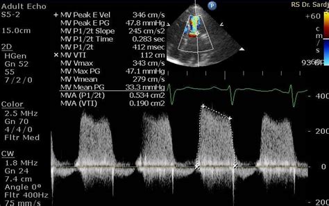
lv volume mitral stenosis | severe mitral stenosis lv volume mitral stenosis Several early studies demonstrated abnormalities of left ventricular (LV) systolic contractility in about 25% of patients with mitral stenosis, with a predilection for the posterior wall. LV ejection is usually mildly decreased. $63.99
0 · severe mitral stenosis mva
1 · severe mitral stenosis
2 · mitral ventricular stenosis
3 · mitral stenosis treatment pdf
4 · mitral stenosis score chart
5 · mitral stenosis normal range
6 · mitral stenosis calcification chart
7 · low graded mitral stenosis
There was a string of lovers, among which the best-known, and probably most influential on her life, was Arthur "Boy" Capel, who died in a car accident in 1919. .
severe mitral stenosis mva
where to buy hermes shoes
Several early studies demonstrated abnormalities of left ventricular (LV) systolic contractility in about 25% of patients with mitral stenosis, with a predilection for the posterior wall. LV ejection is usually mildly decreased. Patients with low‐flow, low‐gradient severe mitral stenosis had higher arterial afterload, greater prevalence of atrial fibrillation, more subvalvular thickening, and decreased .Conversely, when stenosis is severe and valve opening is significantly restricted, the volume of flow across the valve is reduced, leading to reduced filling of the LV and consequently low LV .Mitral stenosis is defined as a narrowing of the mitral valve orifice. There are many causes of mitral stenosis, the most common of which are rheumatic heart disease, congenital .
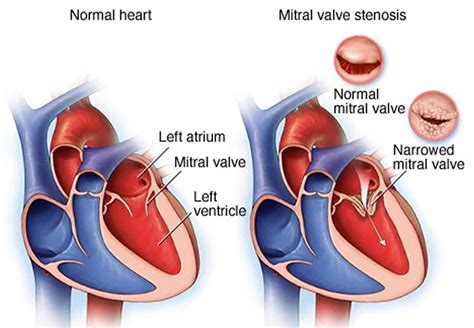
What is mitral valve stenosis? Mitral stenosis is a narrowing of the mitral valve opening. Mitral stenosis restricts blood flow from the left atrium to the left ventricle. Watch a .
The aim of this paper was to detail the recommended approach to the echocardiographic evaluation of valve stenosis, including recommendations for specific measures of stenosis .Pressure–volume relationship before (blue) and after (red) transcatheter aortic valve implantation in a patient with moderate aortic stenosis and depressed left ventricular systolic function. . Coexisting mitral regurgitation is common in patients with mitral stenosis and may elevate the transmitral pressure gradient (due to increased transmitral volume flow rate); however, both 2D echo and T½ valve area .The diastolic volume of the LV is within the low normal limit in all patients; however, the LV filling pressure may be increased in a few patients because of LV scarring and secondary to leftward .
Mitral stenosis (MS) is a form of valvular heart disease. Mitral stenosis is characterized by narrowing of the mitral valve orifice. Today, the most common cause of mitral stenosis is rheumatic fever, but the stenosis usually appears clinically relevant only .
Several early studies demonstrated abnormalities of left ventricular (LV) systolic contractility in about 25% of patients with mitral stenosis, with a predilection for the posterior wall. LV ejection is usually mildly decreased. Patients with low‐flow, low‐gradient severe mitral stenosis had higher arterial afterload, greater prevalence of atrial fibrillation, more subvalvular thickening, and decreased left ventricular compliance compared with those with high‐gradient mitral stenosis.Conversely, when stenosis is severe and valve opening is significantly restricted, the volume of flow across the valve is reduced, leading to reduced filling of the LV and consequently low LV diastolic filling pressures.
severe mitral stenosis
Mitral stenosis is defined as a narrowing of the mitral valve orifice. There are many causes of mitral stenosis, the most common of which are rheumatic heart disease, congenital malformations, radiation complications, metastases, myxoma, cardiac thrombi, etc (Table 1). What is mitral valve stenosis? Mitral stenosis is a narrowing of the mitral valve opening. Mitral stenosis restricts blood flow from the left atrium to the left ventricle. Watch a mitral valve stenosis animation. What problems can result .The aim of this paper was to detail the recommended approach to the echocardiographic evaluation of valve stenosis, including recommendations for specific measures of stenosis severity, details of data acquisition and measurement, and grading of severity.Pressure–volume relationship before (blue) and after (red) transcatheter aortic valve implantation in a patient with moderate aortic stenosis and depressed left ventricular systolic function. Contractility increases and the left ventricular is unloaded as characterized by a left shift of the pressure–volume loop.
Coexisting mitral regurgitation is common in patients with mitral stenosis and may elevate the transmitral pressure gradient (due to increased transmitral volume flow rate); however, both 2D echo and T½ valve area measurements remain accurate.
The diastolic volume of the LV is within the low normal limit in all patients; however, the LV filling pressure may be increased in a few patients because of LV scarring and secondary to leftward shifting of inter-ventricular septum due to early filling of RV and RV distension. Mitral stenosis (MS) is a form of valvular heart disease. Mitral stenosis is characterized by narrowing of the mitral valve orifice. Today, the most common cause of mitral stenosis is rheumatic fever, but the stenosis usually appears clinically relevant only .
Several early studies demonstrated abnormalities of left ventricular (LV) systolic contractility in about 25% of patients with mitral stenosis, with a predilection for the posterior wall. LV ejection is usually mildly decreased. Patients with low‐flow, low‐gradient severe mitral stenosis had higher arterial afterload, greater prevalence of atrial fibrillation, more subvalvular thickening, and decreased left ventricular compliance compared with those with high‐gradient mitral stenosis.Conversely, when stenosis is severe and valve opening is significantly restricted, the volume of flow across the valve is reduced, leading to reduced filling of the LV and consequently low LV diastolic filling pressures.Mitral stenosis is defined as a narrowing of the mitral valve orifice. There are many causes of mitral stenosis, the most common of which are rheumatic heart disease, congenital malformations, radiation complications, metastases, myxoma, cardiac thrombi, etc (Table 1).
What is mitral valve stenosis? Mitral stenosis is a narrowing of the mitral valve opening. Mitral stenosis restricts blood flow from the left atrium to the left ventricle. Watch a mitral valve stenosis animation. What problems can result .The aim of this paper was to detail the recommended approach to the echocardiographic evaluation of valve stenosis, including recommendations for specific measures of stenosis severity, details of data acquisition and measurement, and grading of severity.
Pressure–volume relationship before (blue) and after (red) transcatheter aortic valve implantation in a patient with moderate aortic stenosis and depressed left ventricular systolic function. Contractility increases and the left ventricular is unloaded as characterized by a left shift of the pressure–volume loop. Coexisting mitral regurgitation is common in patients with mitral stenosis and may elevate the transmitral pressure gradient (due to increased transmitral volume flow rate); however, both 2D echo and T½ valve area measurements remain accurate.


$113K+
lv volume mitral stenosis|severe mitral stenosis


